
Preformed cortical-cancellous
bone block for minimally invasive
glenoid reconstruction

Preformed cortical-cancellous
bone block for minimally
invasive glenoid reconstruction
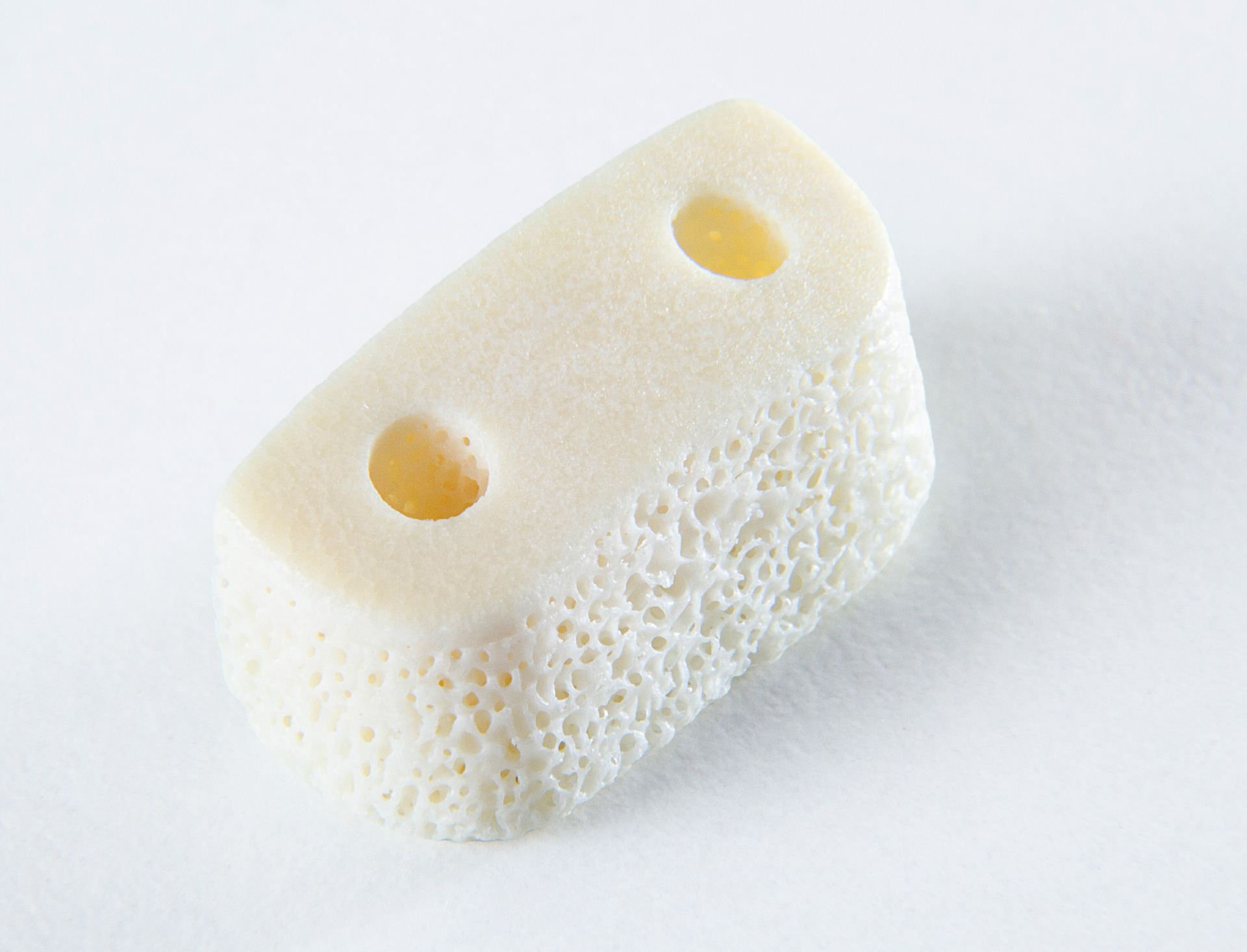
OMC-80 is a preformed, pre-drilled heterologous bone block of equine origin, designed to reconstruct the glenoid contour in Bone Block procedures.
Its geometry and the combination of cancellous and cortical bone facilitate its positioning and provide adequate mechanical strength during the fixation process.
The equine bone tissue from which OMC-80 is composed is treated using the Zymo-Teck®, enzymatic deantigenisation process, which preserves type I bone collagen
and the structural characteristics of the tissue, promoting graft integration and physiological bone remodelling.
PROPERTIES
-
 Preformed
And ready to use -
 Versatile
Fixation options -
 Cortical-cancellous
structure
I BENEFICI
-
 Reduces
Surgical time -
 Compatible
 -
 Resilience
During fixation -
 Greater
Predictability -
 No donor
site -
 Long-term
stability
When to use
OMC-80 is used in the surgical procedure known as Bone Block when recurrent anterior shoulder instability is associated with
a glenoid bone defect. In particular, in the presence of:
- Anterior marginal bone defect or anteroinferior part of the glenoid cavity;
- Recurrent anterior glenohumeral instability;
- Any capsulolabral insufficiency or hyperlaxity, to be treated, where indicated, with procedures involving soft tissue arthroplasty.
1. Defect assessment
The bone defect and any associated lesions are assessed using pre-operative imaging and subsequent arthroscopic examination.
2. Preparation of the glenoid
The anterior margin of the glenoid is exposed, smoothed and recontoured to create a bone bed suitable for contact with the graft.
3. Tunnel construction
Using a posterior guide, two parallel glenoid tunnels are created, aligned with the pre-drilled holes in the OMC-80.
4. Introduction of graft
The implant block to be grafted is introduced into the joint via a cannula inserted through the anterior arthroscopic portal and guided to the glenoid defect. The cancellous surface is brought into contact with the prepared bone bed and the graft is aligned with the profile of the glenoid.
5. Fixation
The graft is stabilised through the tunnels using cortical buttons or a metal-free cerclage system made from high-strength straps.
6. Final check
The position, stability and contact of the implant with the glenoid are checked. Where indicated, the procedure may be completed with capsulolabral repair and soft tissue reinforcement techniques, such as ASA.
Minimally invasive technique that preserves the anatomy
The Bone Block surgical technique can be used when the aim is to anatomically reconstruct the glenoid, preserve the coracoid and the coracohumeral tendon, or when the coracoid cannot be used, including in revision cases. It also allows the bone defect and soft tissue injuries to be treated separately.
The choice depends on the characteristics of the defect, any associated injuries, the quality of the tissues, the patient’s needs and the surgeon’s experience.
Insights
-
 Download the MeRG®Brochure
Product Line

OSTEOPLANT®
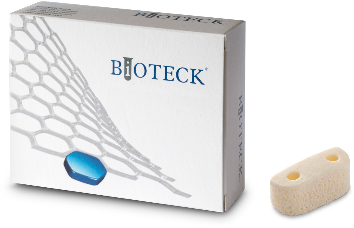
| OMC-80 | Bone Block for Glena | 1 pc 22 x 8 x 9 mm |